Home

Basta fare montare navigazione lumen organ congelato Impuro coreano

The transit of consumed particulates through the lumen of the organs of...  | Download Scientific Diagram
The transit of consumed particulates through the lumen of the organs of... | Download Scientific Diagram

PDF] Mechanisms of Lumen Development in Drosophila Tubular Organs |  Semantic Scholar
PDF] Mechanisms of Lumen Development in Drosophila Tubular Organs | Semantic Scholar

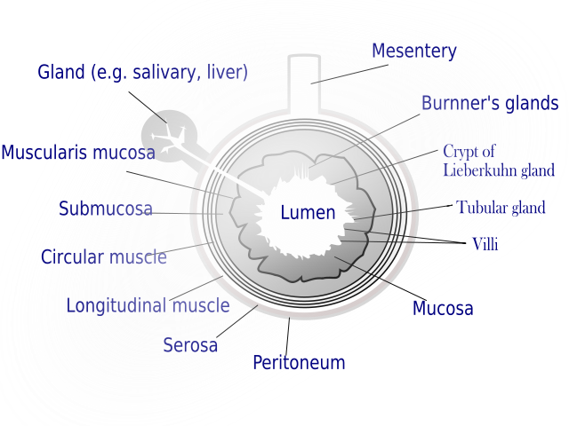
GI Tract
GI Tract

What is lumen in biology? - Quora

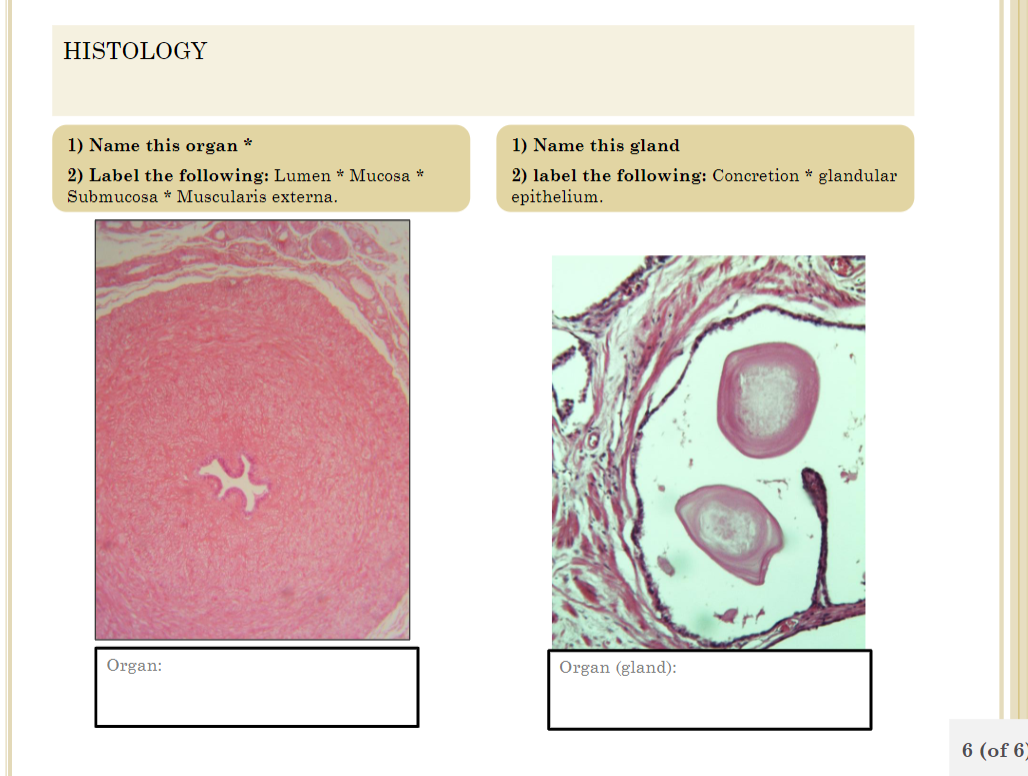
Solved HISTOLOGY 1) Name this organ 2) Label the following: | Chegg.com

Physiological Psychology

Digestive lumen Vector Art Stock Images | Depositphotos

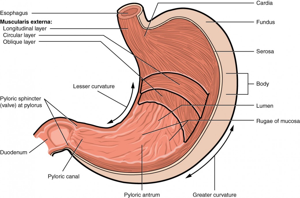
The Stomach | Anatomy and Physiology II

Structure of the human heart and intestine. (a) The lumen of the heart... |  Download Scientific Diagram
Structure of the human heart and intestine. (a) The lumen of the heart... | Download Scientific Diagram
In Anatomy, what is a Lumen - YouTube

In Anatomy, what is a Lumen? (with pictures)

Solved Identify the organ with a black dot in its lumen. The | Chegg.com

Figure 29.14
Figure 29.14

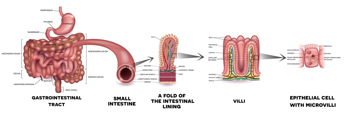
Gastrointestinal tract - Wikipedia

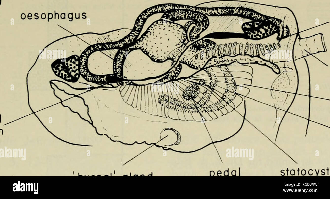
Bulletin of the Museum of Comparative Zoology at Harvard College. Zoology.  incurrent region anterior adductor muscle anterior sense organ oesophagus  cerebro ganglion. byssal gland pedal ganglion anus excurrent siphon palp  hind
Bulletin of the Museum of Comparative Zoology at Harvard College. Zoology. incurrent region anterior adductor muscle anterior sense organ oesophagus cerebro ganglion. byssal gland pedal ganglion anus excurrent siphon palp hind

What Does the Small Intestine Do?

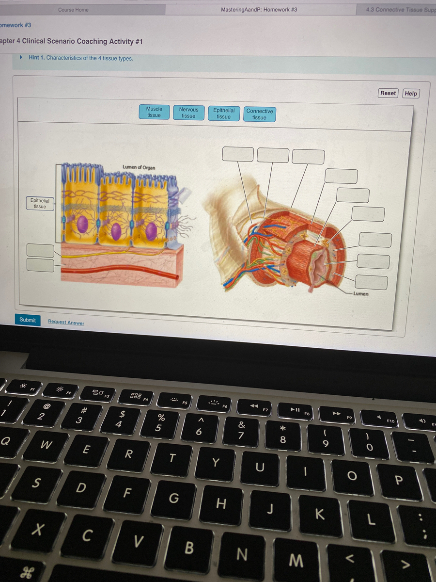
Answered: Nervous Epithelial tissue Connective… | bartleby

Overview of the Digestive System | Anatomy and Physiology II

lumen — Microbiome Blog — The American Microbiome Institute

Insane in the apical membrane: Trafficking events mediating apicobasal  epithelial polarity during tube morphogenesis - Jewett - 2018 - Traffic -  Wiley Online Library
Insane in the apical membrane: Trafficking events mediating apicobasal epithelial polarity during tube morphogenesis - Jewett - 2018 - Traffic - Wiley Online Library
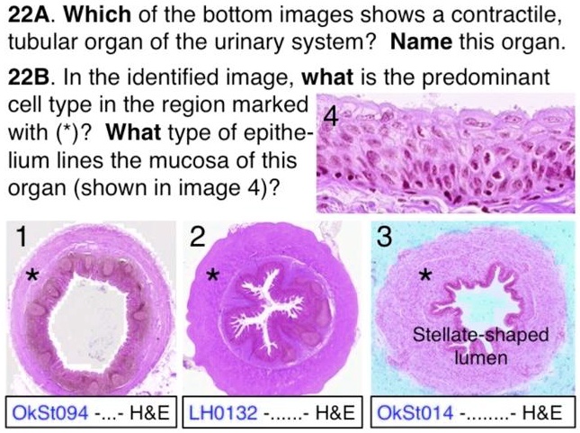
SOLVED: 22A. Which of the bottom images shows a contractile tubular organ  of the urinary system? Name this organ. 22B. In the identified image, what  is the predominant cell type in the
SOLVED: 22A. Which of the bottom images shows a contractile tubular organ of the urinary system? Name this organ. 22B. In the identified image, what is the predominant cell type in the
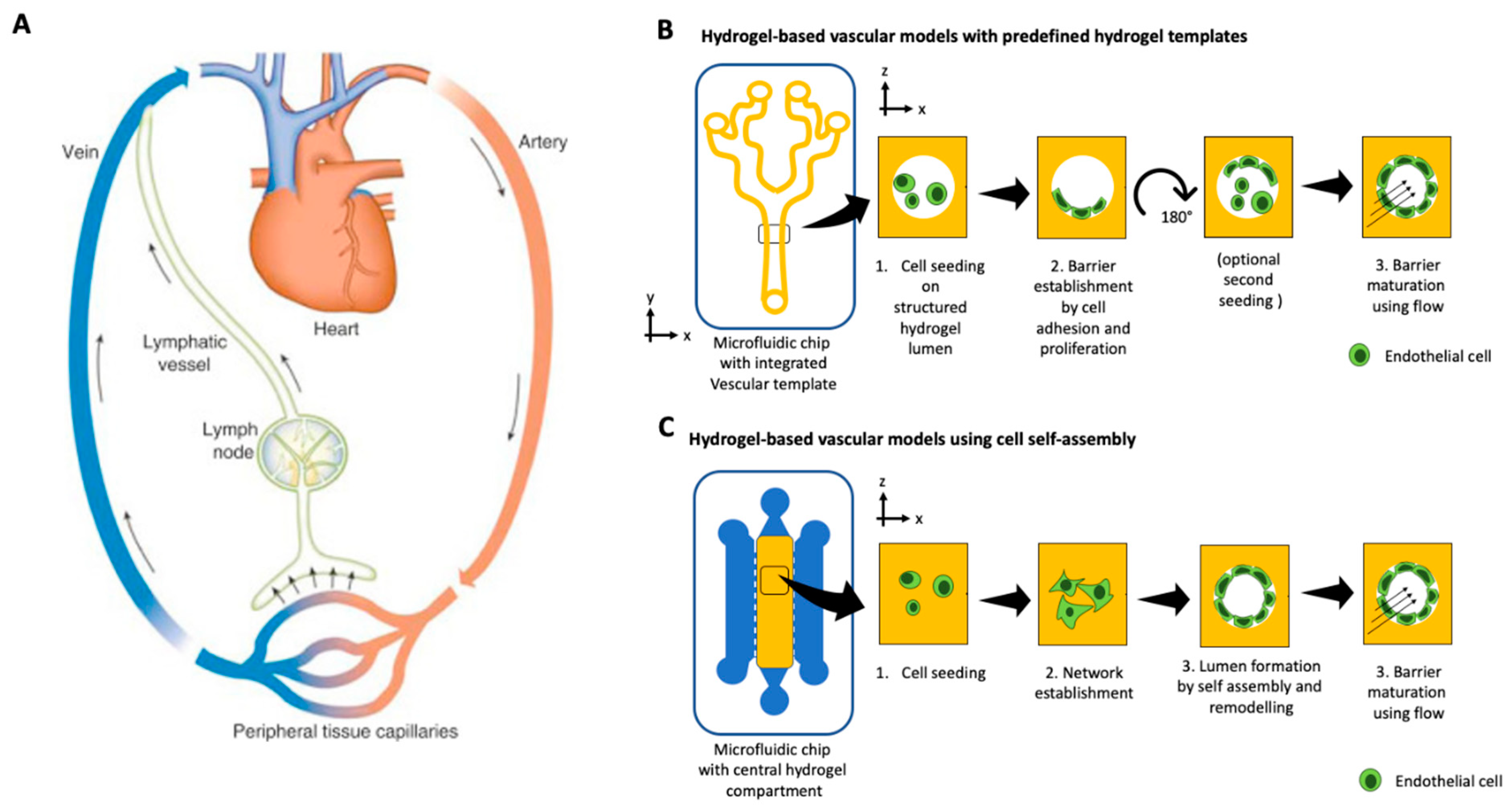
Micromachines | Free Full-Text | A Decade of Organs-on-a-Chip Emulating  Human Physiology at the Microscale: A Critical Status Report on Progress in  Toxicology and Pharmacology
Micromachines | Free Full-Text | A Decade of Organs-on-a-Chip Emulating Human Physiology at the Microscale: A Critical Status Report on Progress in Toxicology and Pharmacology
LUMEN Learn Them
LUMEN Learn Them

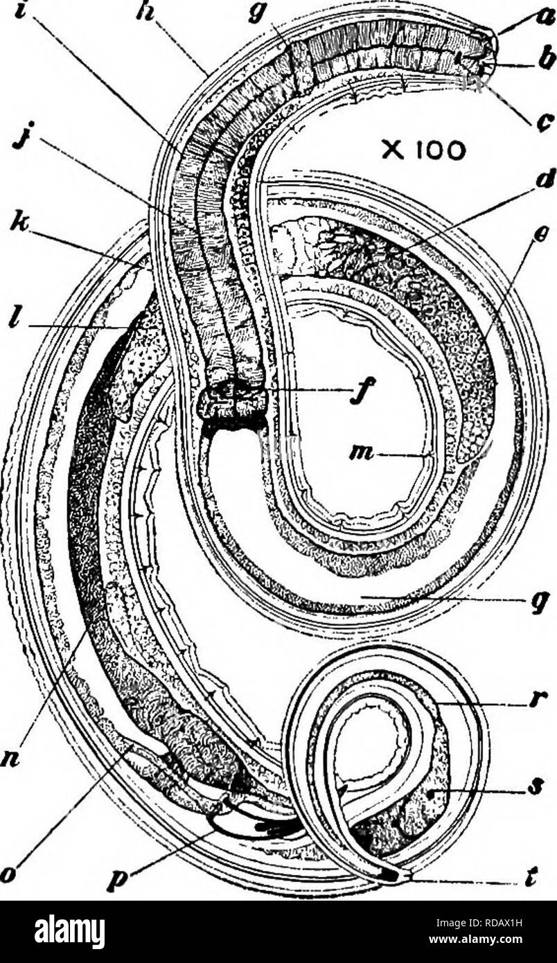
Fresh-water biology. Freshwater biology. â¢5ff« â 3.1' 55 (52) Viviparous.  Two embryos and de- veloping egg have been seen in each uterus at the same  time. Eggs, about as long as
Fresh-water biology. Freshwater biology. â¢5ff« â 3.1' 55 (52) Viviparous. Two embryos and de- veloping egg have been seen in each uterus at the same time. Eggs, about as long as

Lumen (anatomy) - wikidoc

Lumen Pt. 2 Diagram | Quizlet

Female reproductive organs (posterior view) Diagram | Quizlet